Advanced Diagnostics
Latest technologies help find answers
At Tauber Eye Center, we are committed to do all we can to accurately diagnose eye problems and deliver state-of-the-art treatment and care for our patients. We have invested substantially in the latest technological tools to help us accomplish this goal. Eye care providers in the Midwest region often refer patients to us specifically because of the advanced tools we have in our practice.

DRY EYE DIAGNOSIS
Do you really have dry eye? Are there multiple causes of dry eye symptoms. There certainly are, and precise identification of the abnormality driving each patient’s symptoms is the way to achieve resolution of complaints. The symptoms of dry eyes are very similar to other conditions such as blepharitis (eyelid inflammation) and eye allergies.
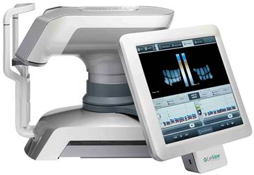
Osmolarity is a measure of how dilute or dense tears are, and has been shown to be the most sensitive and specific measure of dry eyes. We use the newly developed Tearlab device to measure tear osmolarity in all dry eye patients. We also use sophisticated infrared photography to look at the oil producing glands in the eyelids, as abnormalities of this tear component are the root cause of dry eye in almost 70% of patients. We use both the TearScience LipiView device and also the Oculus Keratograph 5 to examine these glands.
CONFOCAL MICROSCOPY
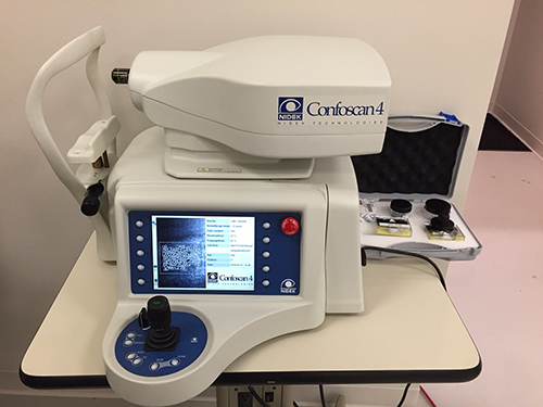
The cornea, the clear “window” that is the front of the eye, can develop serious infections, from bacteria, viruses, fungus, mold, yeast or even parasites. Precise identification of the germ responsible is necessary to select proper medical treatment. The confocal microscope provides highly magnified imaging of the cornea in a slice-by-slice manner to visualize the type of infection that is present. We can also evaluate and follow the health of corneal transplants with serial microscopic photographs. This advanced microscope is one of very few in the Kansas City region.
SD-OCT ADVANCED IMAGING
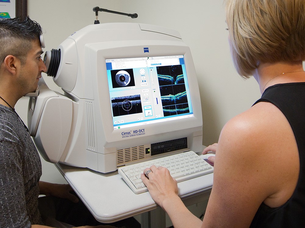
Optical Coherence Tomography (OCT) is a non-contact medical imaging technology similar to ultrasound and MRI. With OCT, reflected light produces detailed cross-sectional and 3D images of the eye. Earlier versions of this tool (TD-SDT) provided 400 scans per second. The newer Spectral-domain OCT (SD-OCT) uses a significantly faster, non-mechanical technology. SD-OCT simultaneously measures multiple wavelengths of reflected light across a spectrum, and is 100 times faster than TD-OCT and acquires 40,000 A-scans per second. The increased speed and number of scans translates into higher resolution and a better chance of observing disease. We use this imaging tool on the cornea, retina and to study the health of the optic nerve.
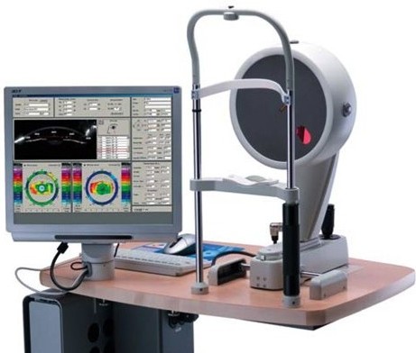
PENTACAM CORNEAL IMAGING
Most corneal specialists make frequent use of topography, an imaging method that shows details about the curvature (shape) of the corneal surface, that is the primary place where light is refracted (bent) to focus within the eye. We use a state-of-the-art camera for our imaging. The Pentacam is a high-resolution rotating Scheimpflug camera system for anterior segment analysis. It provides crisp images of cornea, iris and crystalline lens. It measures the anterior and posterior corneal topography and elevation, total corneal refractive power, corneal power distribution, automatic chamber angle measurement in 360°, chamber depth and volume, and shows corneal and crystalline lens optical opacities. Special software allows for early detection of keratoconus, a progressive condition that affects the cornea.